
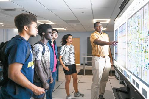
Super-Resolve Imaging Using AI May Help Detect Disease Early
Dr. Kofi Deh, a researcher in the Howard’s Department of Physics and Astronomy is collaborating with the Molecular Imaging Laboratory to focus on imaging changes in metabolism — the chemical processes that allow cells to function. These changes can be one of the first indicators of cancer, diabetes, neurological disorders, and a range of other diseases, often occurring long before structural cell changes begin. However, current imaging methods like PET scans and MRI present several challenges, including an inability to see the entire metabolic process, low specificity, and cost. As the models used for image quality improvement and analysis further improve, artificial intelligence will become more critical to quickly and accurately identifying abnormalities and metabolic changes, leading to faster, more precise diagnoses and treatment.
Deh’s research improves image visual quality by using an artificial neural network to “super-resolve” scans. This requires both natural high-quality images and data on the physical processes being imaged.
“To acquire the higher resolution images that you need for training the neural network, you have to resort to physics-informed methods, where you actually model the processes you are trying to super-resolve,” Deh said.
Deh gave the example of measuring diffusion — the gradual spreading out of molecules from areas of high concentration to low concentration — within human brains.
“You cannot measure diffusion at each point in the brain,” he explained. “What you can do, though, is measure pressure at several points in the brain, or at several arteries. And when you do that you can set up a number of equations which model blood flow.”
These equations can then be used, along with anatomical images, by the neural network to help ensure images are enhanced accurately.
Keep Reading
-
 Epiphanies, Discovery, and Research
New Programs in AI, Microchip Design, and Construction Engineering Management Aggressively Prepare Howard Students for Fields Driven By Evolving Technologies
Jun 22, 2026 4 minutes -
 Artificial Intelligence
Demystifying AI: The College of Engineering and Architecture’s AI Tinkery Series Advances Practical AI Fluency
Jun 22, 2026 4 minutes -
 Culture Creators: Arts and Entertainment
Howard University Professor and Jazz Musician Marcus Johnson Delivers Wisdom on Legacy, Entrepreneurship, AI at 2026 Essence Festival of Culture
Jul 10, 2026 7 minutes